Research News
-
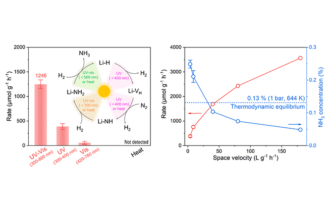 07 01, 2026Researchers Unlock Wavelength-Dependent Stepwise Nitrogen Fixation and Hydrogenation to Break Thermodynamic Limits in Ammonia SynthesisA research team led by Prof. CHEN Ping and Prof. GUO Jianping from the Dalian Institute of Chemical Physics of the Chinese Academy of Sciences, in collaboration with Prof. WU Anan from Xiamen University, revealed a wavelength-dependent photo-driven pathway for ammonia synthesis over lithium hydride (LiH), enabling the decoupling of two long-standing conflicting reaction steps in thermal catalysis.Ammonia is irreplaceable for agricultural fertilizers and is emerging as a zero-carbon energy carriers. Yet the dominant industrial Haber-Bosch process operates under extreme conditions—temperaures above 400 °C and pressure exceeding 100 bar—consuming large amounts of fossil energy and releasing significant carbon emissions.The fundamental limitation lies in the universal scaling relation of thermal catalysis: catalysts capable of activating inert N≡N bonds typically bind nitrogen intermediates too strongly for subsequent hydrogenation, whereas weak nitrogen adsorption impedes N2 activationRecently, a research team led by Prof. CHEN Ping and Prof. GUO Jianping from the Dalian Institute of Chemical Physics of the Chinese Academy of Sciences, in collaboration with Prof. WU Anan from Xiamen University, revealed a wavelength-dependent photo-driven pathway for ammonia synthesis over lithium hydride (LiH), enabling the decoupling of two long-standing conflicting reaction steps in thermal catalysis.This study was published in Journal of the American Chemical Society.The researchers identified a two-stage, wavelength-separated catalytic mechanism. Ultraviolet (UV) light (300-400 nm) exclusively activates LiH to facilitate N2 dissociation and formation of nitrogen intermediates, while both UV and visible light activate Li2NH/LiNH2 species to lower hydrogenation barriers, promote NH3 release, and regenerate LiH.Under 1 bar and 644 K, the combined UV-visible illumination yields an ammonia concentration of 0.25% at the reactor outlet, nearly twice the thermal equilibrium limit (0.13%). The corresponding ammonia production rate reaches 1,246 μmol g-1 h-1, significantly outperforming either UV- or visible-only irradiation.Density functional theory (DFT) calculations confirm that wavelength-specific photoexcitation selectively reshapes reaction energy barriers, thereby breaking the restrictive scaling relations of thermal ammonia synthesis.Schematics for the catalytic performance and mechanism of photo-driven ammonia synthesis process mediated by LiH(Image by GUAN Yeqin)"Our study introduces a wavelength-dependent photoexcitation strategy that independently regulates different catalytic steps via tailored light wavelengths, offering a new guidance for mild, solar-driven nitrogen fixation and other energy-intensive catalytic reactions," said Prof. CHEN.
 -
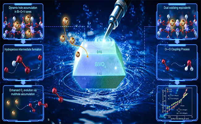 06 25, 2026Scientists Visualize Nanoscale Dynamic Multihole-Mediated Water Oxidation on Single-Particle CatalystsA research team led by Prof. LI Can and Prof. FAN Fengtao from the Dalian Institute of Chemical Physics (DICP) of the Chinese Academy of Sciences (CAS), in collaboration with Prof. LI Jian-feng's team from Xiamen University integrated operando electrochemical shell-isolated nanoparticle-enhanced Raman spectroscopy (SHINERS) with nanoscale electrochemical reaction imaging to spatially resolve the atomic-scale interplay between hole transfer dynamics and the evolution of water oxidation intermediates on faceted BiVO4 particles.Water oxidation reaction, widely recognized as the kinetic bottleneck of artificial photosynthesis, limits solar fuel efficiency. It demands the dynamic transfer of multiple electrons and protons at a complex catalyst–liquid interface, where photogenerated holes dynamic accumulate on the catalyst surface, drive atomic-scale structural rearrangements, and regulate chemical bond breaking and formation. Despite its status as a "Holy Grail" reaction for renewable solar fuels, the dynamic spatial coupling of charge transfer, localized structural motifs and active-site evolution remains unresolved in space and time, particularly as identified under operando conditions, obscuring key mechanistic pathways.Now, a research team led by Prof. LI Can and Prof. FAN Fengtao from the Dalian Institute of Chemical Physics (DICP) of the Chinese Academy of Sciences (CAS), in collaboration with Prof. LI Jian-feng's team from Xiamen University, has broken this barrier. In a paper published in Nature Nanotechnology, they integrated operando electrochemical shell-isolated nanoparticle-enhanced Raman spectroscopy (SHINERS) with nanoscale electrochemical reaction imaging to spatially resolve the atomic-scale interplay between hole transfer dynamics and the evolution of water oxidation intermediates on faceted BiVO4 particles.The researchers discovered a critical hole density threshold that dictates pathway bifurcation. Below a surface hole density of 0.67 nm-2, both the (110) and (010) facets operate under single-hole-transfer-limited kinetics, stabilizing hydroperoxo (*OOH) and peroxo (*OO) intermediates. In this regime, the (110) facet exhibits slightly higher intrinsic activity. Above this critical threshold, the (010) facet becomes catalytically superior, exhibiting third-order power-law kinetics driven by dynamic multi-hole accumulation within Bi-O-V core structures via peroxo intermediates. The (110) facet, meanwhile, shifts toward accumulating dual oxidizing equivalents, which facilitates intramolecular O–O coupling but demands higher energy input.These findings shift the current understanding of water oxidation catalysis from a static, site-centric model to a dynamic system governed by mulithole-mediated structural adaptability. Holes, the study shows, are not merely charge carriers-they actively reorganize catalytic centers in response to their own accumulation. This provides a new design principle for artificial photosynthesis: tailoring photocharge–catalyst architectures with atomic-scale precision, rather than focusing solely on static material structures."Our operando nanoscale imaging and spectroscopy reveals that water oxidation is not dominated by static active sites, but by a multihole accumulation-driven, self-adaptive mechanism that dynamically reconfigures reaction pathways on different crystal facets," said Prof. FAN. "This shifts catalyst design from optimizing static structures to engineering the dynamic coupling between photogenerated charges and catalyst architecture," said Prof. LI.
 -
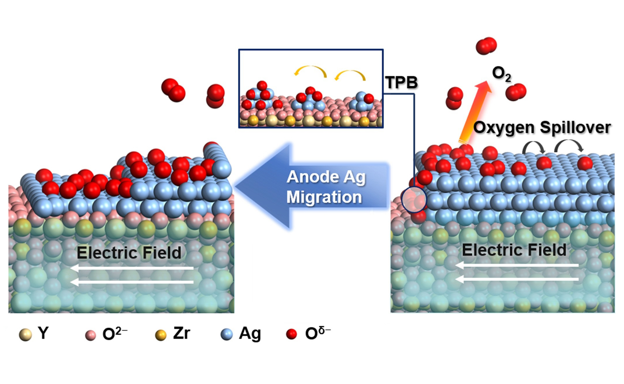 06 24, 2026Electric Field, Oxygen Spillover Team Up Govern Electrode Migration in SOECsA research team led by Prof. FU Qiang from the Dalian Institute of Chemical Physics (DICP) of the Chinese Academy of Sciences (CAS) revealed that the electric field and oxygen spillover operate in a coupled manner to govern electrode migration in SOECs.Dynamic restructuring of electrode surfaces and interfaces often occurs under electrochemical polarization in solid oxide electrolysis cells (SOECs), yet a fundamental understanding of these processes has been hampered by the lack of in situ characterization under realistic operating potential and high temperature.Now, a research team led by Prof. FU Qiang from the Dalian Institute of Chemical Physics (DICP) of the Chinese Academy of Sciences (CAS) found that the electric field and oxygen spillover operate in tandem to govern electrode migration in SOECs.The study was published in Journal of the American Chemical Society.Schematic of Ag migration dynamics in the model SOECs governed by coupled electric field and oxygen spillover (Image by PEI Jinhui)To probe the dynamic behavior, the researchers constructed a planar Ag|yttria-stabilized zirconia (YSZ)|Ag model cell and investigated the dynamic evolution of the working Ag anode using in situ photoemission electron microscopy (PEEM) and micro-region X-ray photoelectron spectroscopy (μ-XPS).They found that the spilled-over oxygen drives Ag transport via the formation of mobile Ag−Oδ− species and the distribution of the electric field dictates the direction and speed of Ag migration. Furthermore, the dynamic restructuring of the Ag anode enhances the oxygen evolution reaction by generating more active triple-phase boundaries (TPBs)."This work establishes an operando methodology for correlating electric-field distributions with oxygen spillover dynamics, enabling a deeper understanding of coupled physical and chemical processes at electrochemical interfaces in high-temperature energy conversion systems," said Prof. FU.
 -
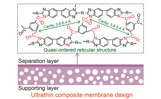 06 23, 2026Perspective: Vanadium Flow Batteries Poised to Power Hundred-Megawatt Long-Duration StorageA research team led by Prof. LI Xianfeng systematically summarize their long-term experience and recent advances in both fundamental research and industrial application of vanadium flow batteries (VFBs).A recent industry perspective article has summarized the long-term experience and recent advances in both fundamental research and industrial application of vanadium flow batteries (VFBs), while charting pathways toward hundred-megawatt-scale long-duration energy storage systems.The perspective, led by Prof. LI Xianfeng from the Dalian Institute of Chemical Physics (DICP) of the Chinese Academy of Sciences (CAS), was published in the newly launched "Down to Business" section of Nature Energy, marking the inaugural article of this column.Design strategies for membranes, flow field, and electrolytes (Image by LU Wenjing)Long-duration energy storage (LDES) is a key technology for integrating high-penetration renewable energy into the power grid and achieving deep decarbonization of power systems. Unlike short duration storage technologies, VFBs offer unique advantages, including high safety, long cycle life, and the decoupling of energy and power capacity, making them a promising candidate for large-scale LDES applications.However, their industrialization still faces several challenges, such as limited stack power density, electrolyte stability issue, high materials costs, and system reliability constraints.In the article, the team systematically reviews their progress in key areas, including critical materials development, stack structure design, and system integration. It further analyzes the key factors influencing the transition from academic research to industrial deployment and charts potential pathways toward hundred-megawatt-scale LDES systems.In recent years, the team has carried out systematic research on core VFBs components, including ion-selectivity and conductive membranes, electrolyte stability, and stack flow-field engineering. Through deep collaboration with industry partners, these advances have been validated and optimized under real operating conditions.By adopting an integrated innovation model spanning fundamental research, pilot-scale validation, and industrial translation, the team has significantly improved the techno-economic performance and engineering readiness of VFB technology.The article also offers a forward-looking perspective on the scalable and low-cost development of VFB systems, delivering valuable insights for the advancement and commercialization of flow battery technologies more broadly.
 -
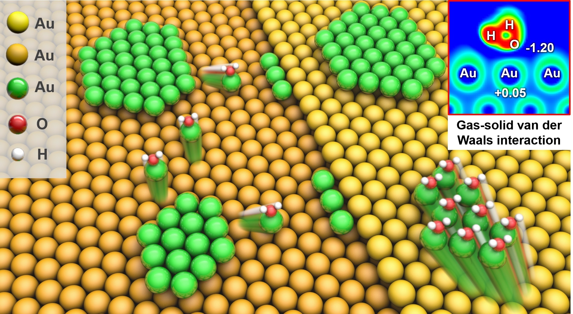 06 23, 2026Gas-Solid van der Waals Interaction Drive Evolution of Surface NanostructuresA research group led by Prof. FU Qiang and Prof. MU Rentao from the Dalian Institute of Chemical Physics (DICP) of the Chinese Academy of Sciences (CAS), in collaboration with Prof. GU Xiangkui from Wuhan University and Prof. LI Weixue from the University of Science and Technology of China, uncovered the dynamic evolution of metal surface nanostructures induced by gas-solid interactions.The gas-induced structural evolution of solid surfaces is a key issue in heterogeneous catalysis and surface science, as it strongly affects catalyst active states, stability, and reaction pathways. However, the direct role of gas-solid physical interactions in driving surface structural reconstruction has remained unclear.Recently, a research group led by Prof. FU Qiang and Prof. MU Rentao from the Dalian Institute of Chemical Physics (DICP) of the Chinese Academy of Sciences (CAS), in collaboration with Prof. GU Xiangkui from Wuhan University and Prof. LI Weixue from the University of Science and Technology of China, uncovered the dynamic evolution of metal surface nanostructures induced by gas-solid interactions. They found that, at the atomic scale, van der Waals interactions between water molecules and surface Au atoms can drive the rapid migration, coalescence, and ripening of Au nanoislands on Au(111) in water vapor.This study was published in the Journal of the American Chemical Society.Schematic of van der Waals interactionsdrive the rapid migration, coalescence, and ripening of Au nanoislands on Au(111) in H2O atmosphere (Image by LIU Changping)The researchers constructed monolayer Au nanoislands on an Au(111) surface and investigated their dynamic evolution under different gas atmospheres using high-pressure scanning tunneling microscopy, near-ambient-pressure X-ray photoelectron spectroscopy, and theoretical calculations.They observed that these Au nanoislands underwent structural evolution — including particle migration, coalescence, and Ostwald ripening — in water vapor at room temperature."This work provides direct atomic-level evidence that gas-solid van der Waals interactions alone can reshape metal surface nanostructures under mild conditions," said Prof. FU. "It challenges the conventional understanding that gas-induced surface reconstruction is mainly governed by strong chemisorption or surface reactions."
 -
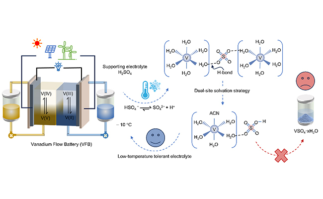 06 15, 2026Scientists Reveal Origin of Low-Temperature Instability in Vanadium Flow Battery ElectrolytesA research team led by Prof. LI Xianfeng from the Dalian Institute of Chemical Physics(DICP), Chinese Academy of Sciences (CAS) uncovered the origin of low-temperature instability in VFB electrolytes and developed a strategy to suppress precipitation through solvation-shell engineering.Vanadium flow batteries (VFBs) hold great promise for large-scale energy storage due to their high safety, long cycle life, and flexible scalability. However, their deployment in cold climate is hindered by the poor low-temperature stability of vanadium electrolytes. In particular, precipitation of divalent vanadium (V(II)) ions in the negative electrolyte can lead to capacity loss and performance degradation, severely limiting the operating temperature range of VFB systems.Recently, a research team led by Prof. LI Xianfeng from the Dalian Institute of Chemical Physics(DICP), Chinese Academy of Sciences (CAS) has uncovered the origin of low-temperature instability in VFB electrolytes and developed a strategy to suppress precipitation via solvation-shell engineering.The study was published in Angewandte Chemie International Edition.By combining single-crystal X-ray diffraction (SCXRD), in situ variable-temperature Raman spectroscopy, and density functional theory (DFT) calculations, the researchers revealed the molecular mechanism of V(II) precipitation.They found that as the temperature drops, the dissociation of HSO4- increase, raising the concentration of SO42- ions in solution. These sulfate anions bridge adjacent [V(H2O)6]2+ complexes through hydrogen-bonding interaction, promoting V(II) dimerization and the formation of ordered clusters that ultimately evolve into VSO4·xH2O precipitates.Low-temperature instability of the negative electrolyte in VFBs and the corresponding suppression strategy (Image by ZHAN Chengbo and LI Tianyu)To tackle this issue, the team developed a dual-site solvation engineering strategy by introducing acetonitrile (ACN) and HCl as co-additives, which simultaneously regulate the first and second solvation shells of V(II) ions. This synergistic approach effectively suppresses ion aggregation and enhances electrolyte stability at low temperatures.As a result, VFBs running on the modified electrolyte maintained an energy efficiency of over 80% for 500 cycles at −10 °C, demonstrating outstanding low-temperature electrochemical performance."Our study provides fundamental insights into the low-temperature behavior of vanadium electrolytes and offers a rational strategy for designing highly stable, wide-temperature-range electrolytes for flow batteries," said Prof. LI.