Recently, in a study published in Angewandte Chemie International Edition, a research team led by Profs. ZHANG Lihua and LIANG Yu from the Dalian Institute of Chemical Physics of the Chinese Academy of Sciences developed a high-throughput strategy for single-cell spatial proteomics boased on an ordered colloidal crystal chromatographic column. This approach improves analytical throughput while maintaining deep proteome coverage, marking a significant advance in single-cell-resolution spatial proteomics.
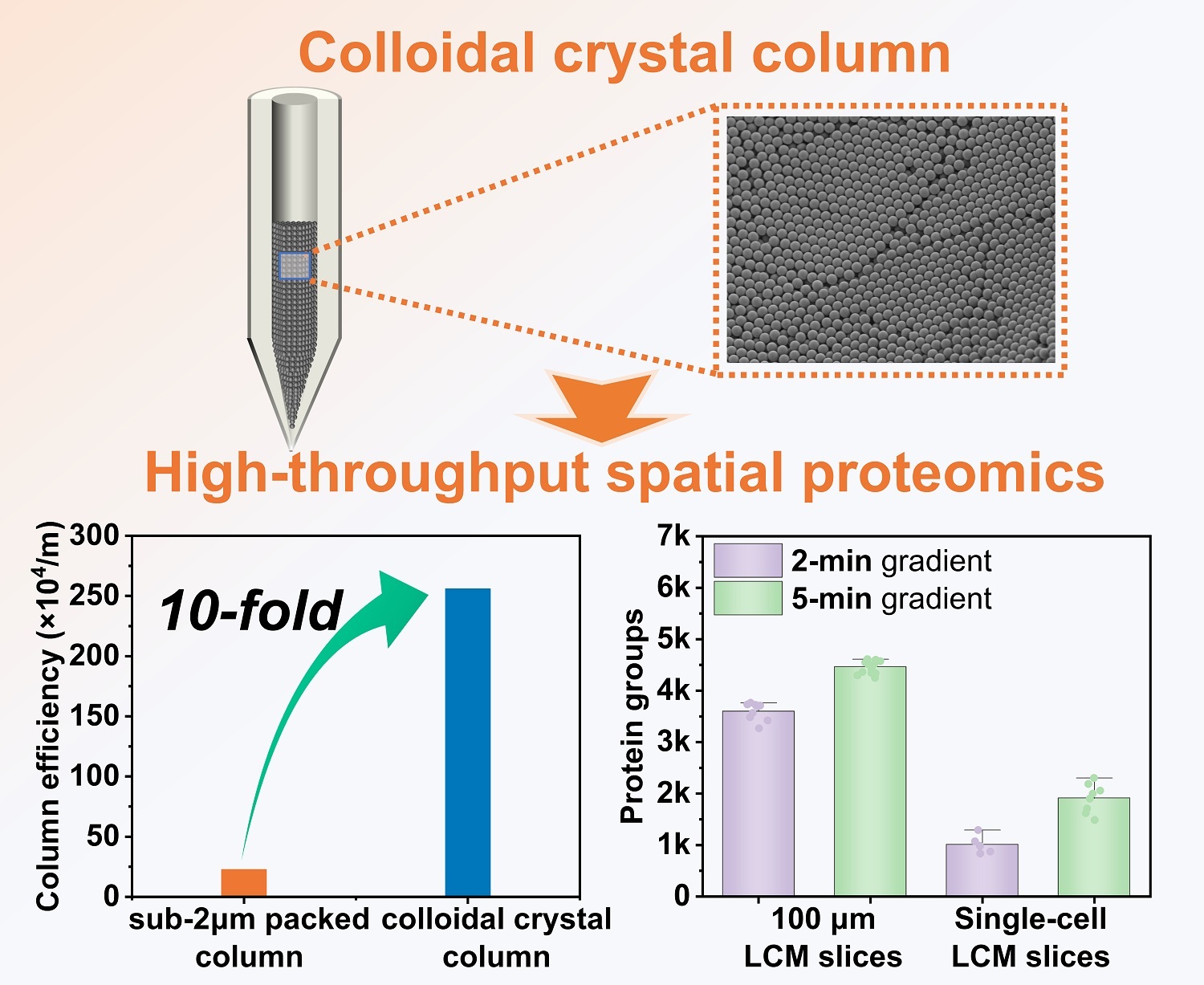
A colloidal crystal chromatographic column assembled from 800 nm monodisperse C18 silica spheres exhibits a tenfold higher column efficiency than conventional sub-2 μm columns, enabling high-throughput spatial proteomics under ultra-short gradients with spatial resolution down to the single-cell level (Image by SUN Haofei and LIANG Yu)
Spatial proteomics enables the characterization of protein distribution within biological tissues and plays a critical role in understanding biological functions and disease mechanisms. Single-cell-resolved spatial proteomics is particularly valuable for investigating cellular heterogeneity, signaling pathways, and intercellular interactions within complex tissue microenvironments.
Currently, mainstream spatial proteomics relies on nanoLC–MS combined with laser capture microdissection (LCM). However, increasing spatial resolution greatly increases the number of tissue slices, creating a severe throughput bottlenecks. Even with advanced chromatographic columns and high-performance mass spectrometry systems, current single-cell analyses typically require about 30 minutes per sample to achieve sufficient proteome coverage—a major speed constraint for large-scale studies.
To address this challenge, the team developed a novel colloidal crystal chromatographic column based on the ordered assembly of 800 nm monodisperse C18 colloidal particles. Benefiting from its highly ordered architecture, submicrometer particle size, and nonporous structure, the column achieved an efficiency exceeding 2 million plates per meter—approximately 10 times higher than that of conventional sub-2 μm chromatographic columns.
This substantial improvement enabled dramatically faster proteomic analysis without compromising proteome depth. Using the ordered colloidal crystal column, the team identified 4,462 ± 119 and 3,597 ± 172 proteins from 100 μm-resolution LCM tissue slices under 5-minute and 2-minute separation gradients, respectively. By comparison, conventional sub-2 μm chromatographic columns required a 30-min gradient to achieve similar proteome coverage.
When applied to single-cell-resolution spatial proteomics, the method delivered exceptional performance, enabling the identification of up to 2,304 proteins from a single hepatocyte slice within only 5 minutes, while still identifying more than 1,000 proteins under a 2-min gradient.
The researchers further applied the method to hepatocellular carcinoma tissues, enabling rapid spatial proteomic characterization of the liver region, early-stage hepatocellular carcinoma, and advanced tumor regions. The approach also revealed proteomic heterogeneity among distinct cell populations within tumor regions at single-cell resolution.
"Our study provides a high-throughput technological tool for single-cell spatial proteomics, molecular atlas construction, and investigations of disease tissue microenvironments," said Prof. ZHANG.