In a recent study published in Journal of the American Chemical Society, a joint research team led by Prof. WANG Fangjun and Prof. XIAO Chunlei from the Dalian Institute of Chemical Physics (DICP) of the Chinese Academy of Sciences, in collaboration with Prof. HANG Wei from Xiamen University and Prof. YIN Zhibin from Shenzhen Institute of Advanced Light Source Facilities, has developed a novel high-spatial-resolution mass spectrometry imaging (MSI) instrument using a single-mode fiber image relay. The system delivers a spatial resolution of 800 nm, allowing researchers to resolve lipid metabolism heterogeneity during drug-induced cell apoptosis at the single-cell level.
Dysregulation of apoptosis represents a hallmark of tumorigenesis, and various anti-cancer drugs induce tumor cell apoptosis via interaction with the cell membrane. During this process, changes in the cell membrane microenvironment and metabolic reprogramming can be characterized by lipidomics. However, due to the small size of single cells and the compositional complexity of lipids, traditional liquid chromatography-mass spectrometry (LC-MS) techniques typically provide only bulk-averaged information, making it difficult to resolve lipid heterogeneity at the single-cell level.
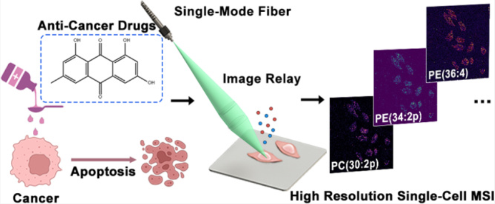
Single-mode fiber image relay mass spectrometry imaging reveals lipid heterogeneity during drug-induced apoptosis (Image by LENG Yixin)
To overcome these limitations, researchers proposed an integrated strategy combining instrument development and MSI methodology. They exploited the mode-filtering properties of single-mode fibers and combined them with a custom-designed optical image relay system to develop a novel high-spatial-resolution laser desorption/ionization source.
The device achieved a spatial resolution of approximately 800 nm while maintaining a long working distance (>25 mm), which helped minimize contamination of the fiber probe. Building on this platform, researchers developed a reflectron time-of-flight mass spectrometer with a mass resolving power greater than 10,000, enabling high spatial resolution MSI of mouse brain tissue sections as well as single cells.
Moreover, researchers integrated high-resolution MSI, cell morphology analysis, and LC-MS to establish a novel approach capable of simultaneously obtaining bulk composition information and single-cell, phenotype-specific lipidomic data. They observed that drug-induced lipid metabolic changes exhibited clear dose- and time-dependent behavior by using this method to study apoptosis in HeLa and HepG2 cells induced by different drugs.
In addition, researchers constructed drug-specific single-cell lipid fingerprints in a multidrug treatment model, revealing metabolic differences associated with distinct pharmacological mechanisms.